Understanding the Anatomy of the Hand
Our hands are a marvel of functionality and comprise 27 bones, 27 joints, 34 muscles, and many ligaments, tendons, nerves, and blood vessels. These structures work together to enable the complex movements we rely on every day—from driving to writing, lifting, and cooking. Understanding the hand anatomy is essential to learning ways to take better care of your hands and prevent injuries.
Key Components of the Hand
Bones & Joints: The Foundation of Movement
Each hand consists of various bones and joints, working together to allow flexibility and precision.
- 8 carpal bones, which connect to the radius and ulna of the forearm, form the wrist.
- The palm consists of 5 metacarpal bones, each connecting to a finger at the metacarpophalangeal (MCP) joint—commonly known as the knuckle joint.
- Fingers and thumbs are made up of phalanges:
- Fingers have three phalanges separated by two interphalangeal joints (PIP and DIP joints).
- The thumb has two phalanges and one interphalangeal joint, providing a wider range of motion.
These bones and joints form a hinge-like structure, allowing your fingers to bend, straighten, and grasp objects with precision.
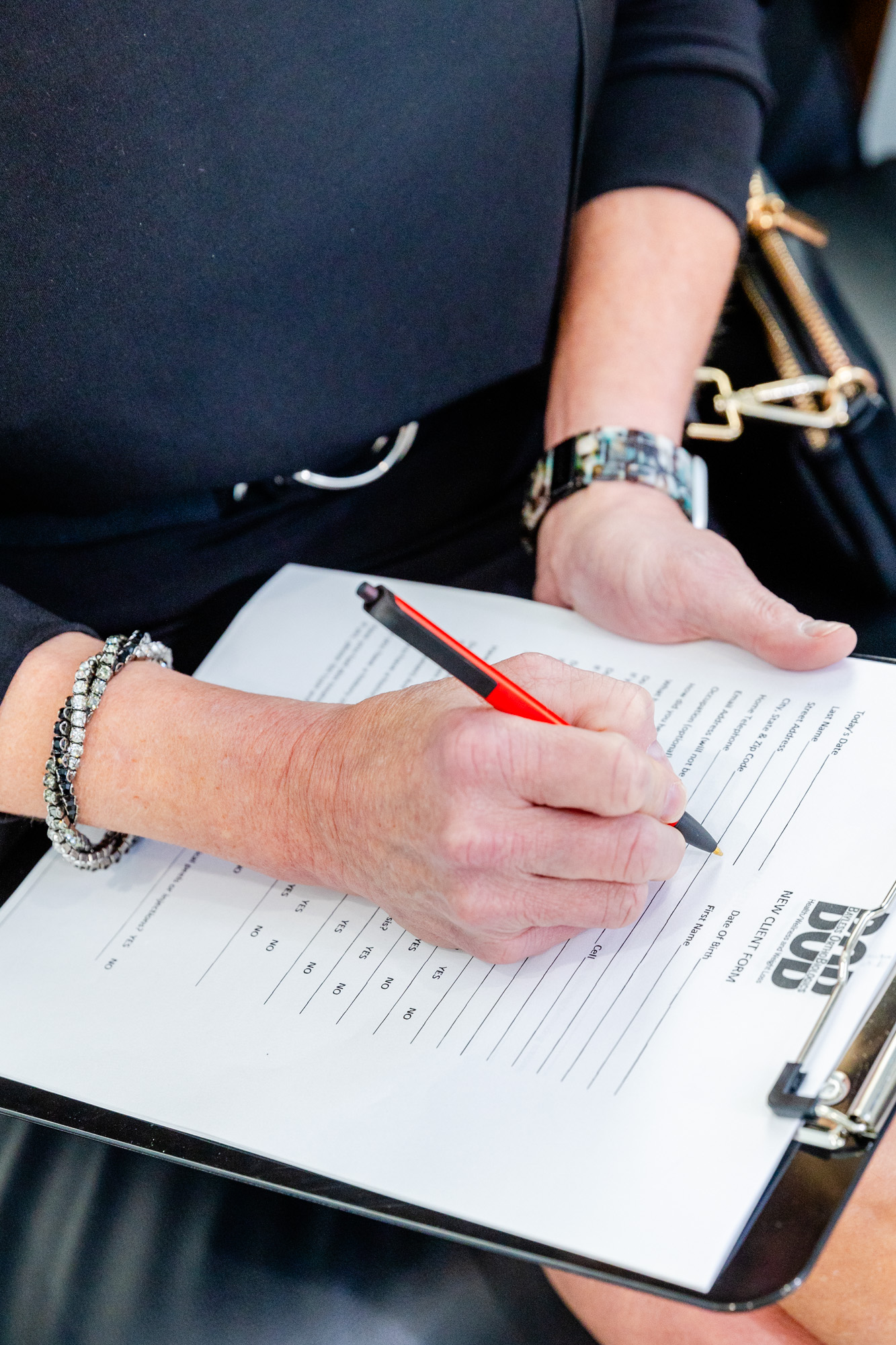
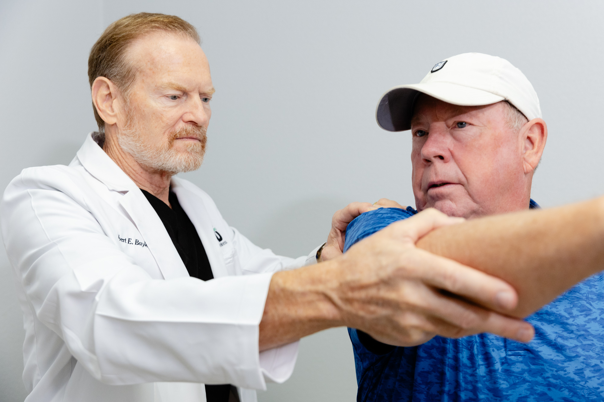
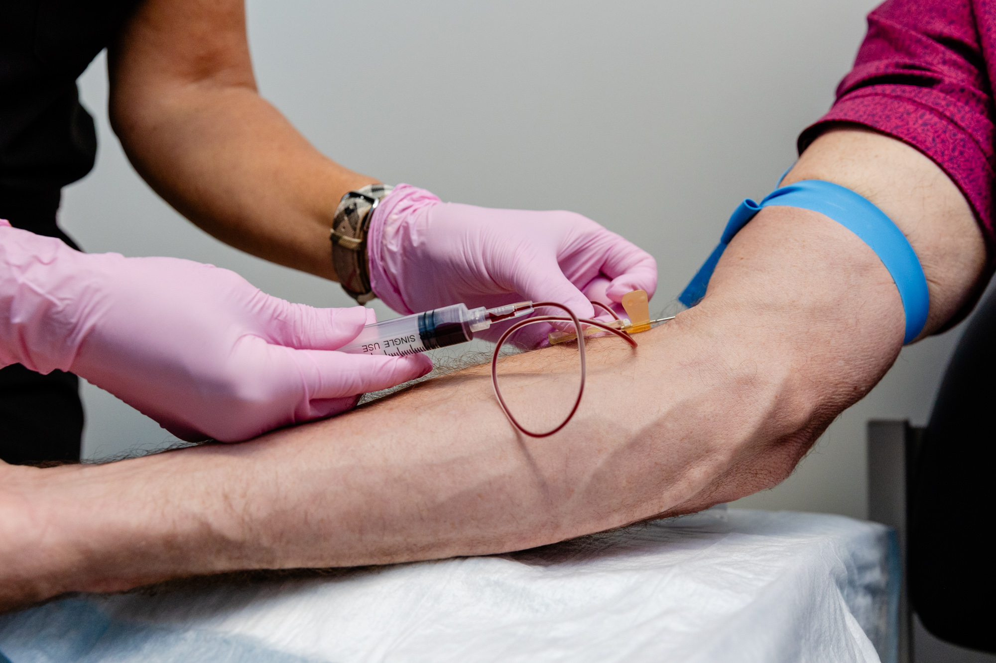
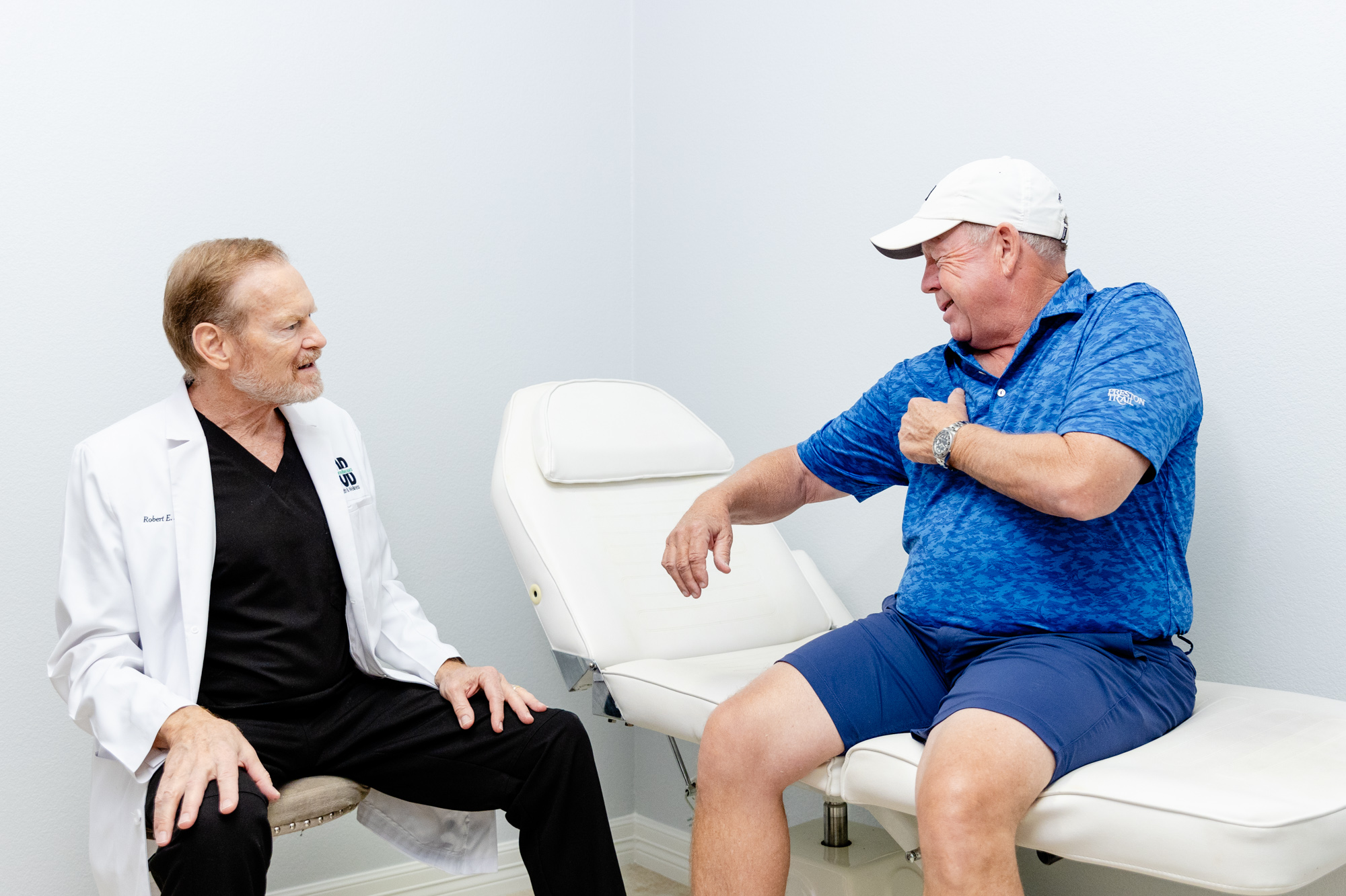
Our Process
Soft Tissues: Stability and Protection
To ensure smooth, pain-free movement, your hands rely on a system of ligaments, tendons, and cartilage for support and cushioning.
- Articular cartilage lines each joint, acting as a shock absorber to prevent bone-on-bone friction.
- Ligaments connect bones to other bones, providing stability and preventing excessive movement.
- Each finger joint has two collateral ligaments, which prevent side-to-side bending.
- The volar plate is the strongest ligament in the hand, stopping the PIP joint from overextending.
Together, these structures stabilize the hand, reducing strain and minimizing injury risk.
Muscles & Tendons: The Power Behind Every Motion
Your hand contains two types of muscles that control movement:
- Intrinsic Muscles: Small muscles within the hand that enable fine motor movements, such as writing or playing an instrument.
- Extrinsic Muscles: Larger muscles that originate in the forearm and elbow, responsible for stronger grip and wrist movement.
Each finger has six muscles controlling movement, helping you perform everything from delicate tasks to powerful grips.
Tendons connect these muscles to bones, allowing them to bend and straighten your fingers:
- Flexor tendons (on the palm side) help bend your fingers.
- Extensor tendons (on the back of the hand) help straighten them.
Healthy tendons are essential for fluid hand movement and injury prevention.
Nerves: Sending Signals for Sensation & Control
The hand’s nerves transmit signals between the brain and muscles, controlling movement and sensation.
- Ulnar Nerve: Travels through Guyon’s canal and controls sensation in the little finger and part of the ring finger.
- Median Nerve: Passes through the carpal tunnel, providing sensation to the palm, thumb, index, middle, and part of the ring finger.
- Radial Nerve: Runs along the thumb side, controlling the back of the hand from the thumb to the middle finger.
Nerve compression or injury can cause tingling, numbness, or weakness, often requiring specialized treatment.
Blood Flow: Circulation for Healing & Strength
Blood vessels travel alongside nerves, delivering oxygen and nutrients to keep your hand strong and functional.
- The Ulnar Artery runs alongside the ulnar nerve, supplying blood to the little and ring fingers.
- The Radial Artery is the largest artery in the hand, running near the thumb, where your pulse often gets measured.
- Additional blood vessels supply the back of the hand, ensuring proper circulation to the fingers and palm.
Proper circulation is essential for healing, especially after an injury.
Bursae: Reducing Friction & Protecting Joints
Bursae are fluid-filled sacs that reduce friction between tendons, bones, and skin. They contain synovial fluid, which acts as a natural lubricant, allowing smooth motion with minimal wear and tear.